
Arbor Vitae: The Neuroanatomist’s Perspective
Dr. Justin Sipla teaches neuroanatomy and neuroscience to medical and graduate students at the University of Iowa. We asked him to ponder how the patterning of a cerebellum’s arbor vitae mirrors Anna Atkins’ cyanotype of a fern below.
A special thank you to the University of Iowa Stanley Museum for collaborating with us on this project and providing us with this image.
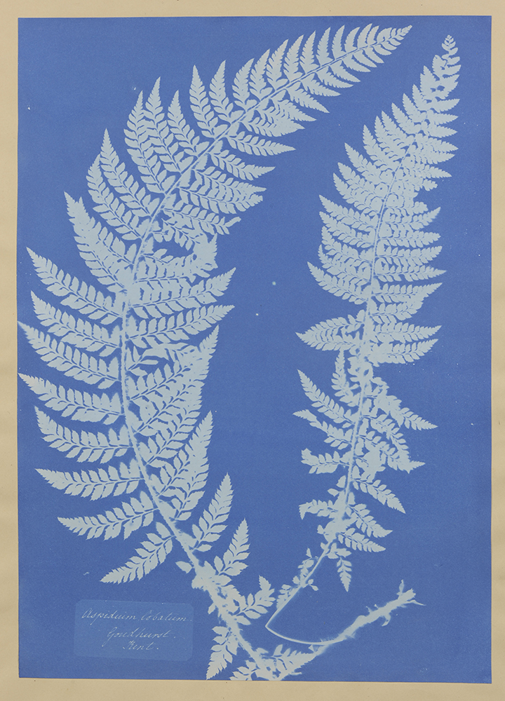
“Aspidium lobatum, Goudhurry, Kent,” 1851–1854
Cyanotype
13 3/4 x 9 5/8 in. (34.93 x 24.45 cm)
University of Iowa Stanley Museum of Art
Museum purchase, 1987.3
I watched a Netflix documentary recently about a guy who befriended an octopus while diving off the Cape of Storms (My Octopus Teacher). For me, by far the most moving footage was a moment when the octopus started “playing” with a school of fish, moving the cloud of animals this way and that with its eight tentacles, in apparent delight of the shifting patterns.
All brains really seem to do is pattern the universe, and, using whatever patterns have been established, make predictions about future patterns. That’s basically it. What is it about an encounter with symmetry that sets our brains on fire, whether octopus, human, or (I expect) otherwise?
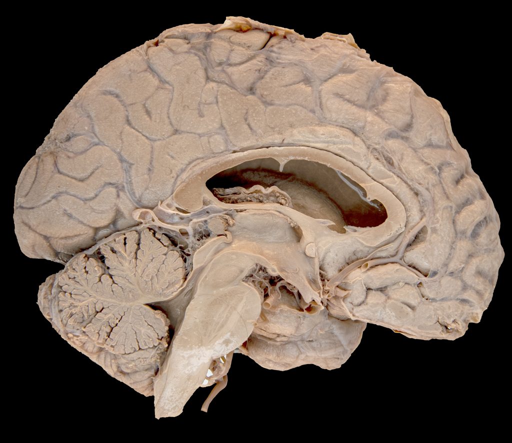
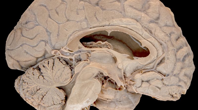
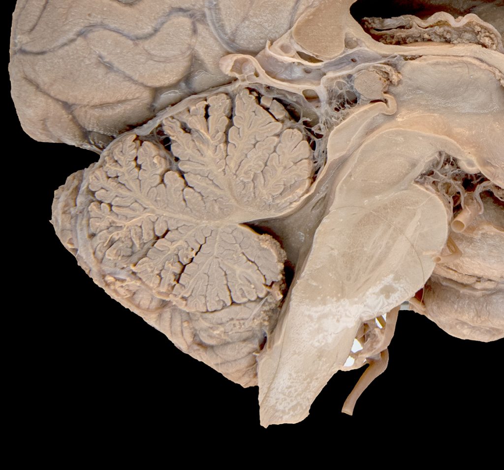
Certainly, the white matter of the cerebellum, or arbor vitae, in cross section resembles a fern, or maybe it’s better to say that a fern resembles a cerebellum. Folded cerebellar white matter is well established in cartilaginous fish, and these first appeared on the evolutionary stage some 400 million years ago in the Devonian, roughly the same time as ferns. Neither came first by much.
Most fern fronds get their shape via circinate vernation, in which new growths uncoil from a fractal spiral. The accordion-like folds of the arbor vitae come about differently. During a tightly choreographed set of developmental events, the surface area of the cerebellar cortex far outgrows the area needed to surround the output nuclei of the deep white matter. Either the cortical sheet folds extensively, or you simply can’t pack enough neurons in there to work the cerebellum’s magic. Gyrification, not circination.
Structural similarities are not merely an aside. Does something even resemble something else unless a brain says it does? When you think about it, all human knowledge is really based on metaphor, brains deciding that some pattern is “like that” or “not like that in ways.” So the arbor vitae is like a fern, and the fern is like an arbor vitae. Some ferns even get their common names after this likeness, for instance the Arborvitae Fern. Though that’s actually a club moss.
About the Author:
Dr. Justin Sipla is a neuroanatomist and curriculum strand director in the Carver College of Medicine. He teaches extensively about the organization and function of the human brain in Iowa’s MD, PA, DPT, neuroscience Ph.D, and Neurology Residency programs.
The Stanley Museum of Art (est. 1969) is a visual arts institution at the University of Iowa and one of the leading university art collections in the country with 15,500 objects. The new Stanley Museum building will open in 2022, find them at @uistanleymusum on Instagram.